Agrovet Pharmaceuticals
INFECTIOUS RESPIRATORY DISEASES
• Chronic Respiratory Disease (CRD)
• Coryza
• Aspergillosis
• Newcastle Disease (ND)
• Infectious Bronchitis (IB)
• Infectious Laryngotracheitis (ILT)
• Avian Influenza
• Turkey Rhinotracheitis/Swollen Head Syndrome
Chronic respiratory Disease
(CRD) (Airsacculitis)
১ এর পর ২ বাদ
৩,৪,৫ আছে লেশন
Cause
Mycoplasma gallisepticum গালি সেপটিকাম (Mg). and E. coli.
Transmission
transmit the organism through the egg . by contact or by airborne dust or droplets.
Species affected
Chickens and turkeys.
Clinical signs
respiratory distress. lack(loss) of appetite, decreased weight gain and increased feed conversion ratios. sneezing, coughing and general signs of respiratory congestion. drop of egg production between 20-30 % .Mortality low.Pericarditis, peritonitis and perihepatitis , Airsacculitis
Internal lesions
A reddish inflamed trachea and/or cheesy exudate in airsacs, especially in complicated cases. In mild Mg infections, slight mucus in trachea and a cloudy or light froth in the airsacs. swollen sinuses under the eyes.
Diagnosis
blood testing , post-mortem examination and isolating the causative Mg organism .Blood serum testing
Treatment
antibiotics or chemotherapeutics . control by medication or vaccination and eradication of Mg infections. antibiotics such as tylosin .
Infectious Coryza
Cause
bacterium: Hemophilus paragallinarum. প্যারা গালি নেরাম
Transmission
by contact and airborne infected dust particles and via the drinking water. Spread by equipment and personnel
Species affected
Chickens
Clinical signs
inflammation of eyes and nose with foul-smelling discharges, conjunctivitis, sneezing and facial swellings. Feed and water intake is reduced( loss of appetite), loss of weight. Egg production drop. Mortality low.
Typical facial edema
Diagnosis
isolation of the organism.
Treatment and control
Treatment with antibiotics, control: eradication and prevention. Vaccines.
Aspergillosis (Fungal Pneumonia)
Cause
fungus, Aspergillus fumigatus. ফিউমি গেটাস
Transmission
fungus spores from ontaminated litter or contaminated feed.
Species affected
Young chicks. Turkey poults, pheasant chicks, quail chicks, ducklings, and goslings.
Clinical signs
depressed and thirsty. Gasping and rapid breathing . Mortality 5 to 50 %. Gross lesions in lungs and airsacs. Yellow white pinpoint lesions. small yellow-green granular fungus growth.
Diagnosis
identified microscopically or sometimes with the naked eye.
Treatment and control
no treatment. Affected chicks should beremoved and destroyed. Strict hygiene.
Newcastle Disease (ND)
Cause
paramyxovirus. প্যারা মিকজু
Transmission
droppings and respiratory discharge . by infected equipment, trucks, personnel, wild birds or air.
Species affected
Chickens and turkeys.
Clinical signs
high mortality with depression and death in 3 to 5 days as major signs. respiratory distress. Labored breathing with wheezing and gurgling, accompanied by nervous signs, such as paralysis or twisted necks (torticollis) are the main signs. Egg production decrease 30 to 50 %. Egg thin shells and eggs without shells. Haemorrhagic proventriculus
Internal lesions
Inflamed tracheas, pneumonia, and/or froth in the airsacs. Haemorrhagic lesions in the proventriculus and the intestines.
Diagnosis
virus isolation, blood testing.
Treatment and control
no treatment. Vaccination control method.
Infectious Bronchitis (IB)
Cause
Corona-virus.
Transmission
through the airborne route. via the air
Species affected
Only chickens.
Clinical signs
cheesy exudate in the bronchi, asphyxia, severe respiratory distress. In older birds no mortality. Egg production decrease, deformed eggs with wrinkled shells.
Internal lesions
Mucus and redness in tracheas, froth in airsacs in older chickens. In young chicks, a yellow cheesy plug at the tracheal bifurcation.
Diagnosis
post-mortem findings(examination), Isolation of the virus. serum tests.
Treatment and control
no treatment. Secondary bacterial infections: antibiotics. Prevention by vaccination.
Infectious Laryngotracheitis (ILT)
Cause
virus herpes group.
Transmission
by the respiratory route. by contaminated people or equipment (visitors, shoes, clothing, egg boxes, used feeders, waterers, cages, crates etc.).
Species affected
Chickens and pheasants.
Clinical signs
Respiratory distress, sloughed tracheal lining and caseous exudate in larynx and trachea. extreme difficulty breathing and die from suffocation. Mortality 1 %. Conjunctivitis and respiratory sounds (wheezing). Haemorrhagic lesions in trachea. Egg production decrease 10 to 50 %.
Diagnosis
coughing up of blood, Bloody mucus and cheesy exudate in larynx and trachea. by histological examination or virus isolation.
Treatment and control
Prevention by vaccination.
Avian Influenza
Cause
myxovirus.মিকজু
Transmission
Airborne virus particles, droppings, and people-carrying virus on their clothing and equipment.
Species affected
Turkeys and ducks main, chickens, geese, and wild birds.
Clinical signs
Respiratory disease with mortality, drop in egg production. Swelling of the head and neck, swollen sinuses with nasal discharge. Mortality is usually low. proventriculus
Diagnosis
by serological or virological methods (virus isolation).
Treatment and control
no treatment. Antibiotics secondary bacterial infections.
Pneumovirus Infections (Turkey Rhinotracheitis/Swollen Head Syndrome)
Cause
pneumovirus. নিউমু ভাইরাস
Transmission
horizontally by contaminated water, personnel and equipment.
Species affected
Turkeys and chickens.
Clinical signs and lesions
sneezing. Rales and nasal discharge, conjunctivitis, swelling of the infraorbital and submandibular sinuses. drop in egg production, respiratory distress. Morbidity high, mortality vary. swollen head syndrome (SHS). swelling of the periorbital and infraorbital sinuses, torticollis, cerebral disorientation and depression. Marked drop in egg production. At necropsy the lesions seen. oedema in the head, purulent or caseous subcutaneous exudate. Rhinitis, tracheitis and sinusitis. Poliserositis affecting the air sacs and pericardium. swollen and congested kidney, fibrinous exudate in the pleural cavity of lungs ,conjunctivitis and sinusitis.
Diagnosis
by the isolation of the organism . serological methods such as the VN test, IFT and ELISA.
Treatment and control
antibiotics for secondary bacterial infections. vaccines.
NEOPLASTIC DISEASES
• Lympoid Leucosis
• Marek’s Disease
Lymphoid Leucosis (LL, Big Liver Disease, Visceral Leucosis)
Cause
retro (leuco) virus.
Transmission
through eggs. horizontal transmission.
Clinical signs
Visceral tumors main feature. lower egg production. Osteopetrosis. bowed, thickened legs. blood leucosis. myeloid leucemias. listless, pale. (no paralysis). weaken, lose weight and die.
Diagnosis
Histopathological examination.
Treatment and control
No treatment. laboratory detection of infected breeders.
Marek’s Disease (MD, Neurolymphomatosis)
Cause
herpes virus.
Transmission
Main transmission by infected premises, by the oral and respiratory routes. horizontal transmission.
Species affected
The domestic fowl.
Clinical signs
weight loss, paralysis. Mortality 5 to 50 %. Leg paralysis. very small gizzard and intestines. Mortality between 10 and 20 weeks of age.
Diagnosis
tumours in liver, spleen, kidneys, lungs, ovary, muscles, or other tissues(lymphoid leucosis same). nerve involvement. Eye involvement (ocular lymphomatosis). Skin involvement (skin leucosis). histological examination.
Treatment and control
Vaccination.
AVIAN ADENOVIRAL DISEASES
• Inclusion Body Hepatitis (Hydropericardium-Hepatitis Syndrome)
• Egg Drop Syndrome 1976 (EDS ’76)
Inclusion Body Hepatitis (Hydropericardium-Hepatitis Syndrome)
Cause
avian adenovirus (for example the Tipton strain)
Transmission
Egg transmission. Horizontal transmission from bird to bird by contact with droppings.
Species affected
Chickens, turkeys and pheasants.
Clinical signs
inclusion body hepatitis. listless, with ruffled feathers. Mortalityis severe up to 25 %.
Internal lesions
mottled livers, many with pinpoint necrotic and haemorrhagic spots. Pale bone marrow and, infectious anemia, gangrenous dermatitis. pale and swollen kidney. The spleen is quite small (atrophy)(atrophic spleen). When Gumboro disease(Atrophic bursa of fabricius), chickens are immunosuppressed. mottled livers, pale bone marrow and gangrenous dermatitis.
Diagnosis
Typical mottled livers with pinpoint lesions, pale bone marrow and kidneys, small spleen and bursa are good indications of the disease. Histological examination, virus isolation.
Treatment and control
No treatment. Antibiotics for secondary bacterial infection. ensure adequate immunity. Vaccination.
Egg Drop Syndrome 1976 (EDS ’76)
Cause
avian adenovirus (strain BC14, virus 127).
Transmission
through the egg. Horizontal spread through infected litter.
Species
Only chickens.
Clinical signs
drop in egg production, inferior eggshell quality and brown eggs, a loss of shell color. anaemic, transient diarrhoea and sometimes the food intake may be reduced(loss of appetite). No increased mortality. Misformed and soft shelled eggs.
Internal lesions
No internal lesions.
Diagnosis
Clinical signs. Virus isolation and antibody tests.
Treatment and control
no treatment. Vaccination.
MISCELLANEOUS VIRAL DISEASES
• Fowl Pox
• Avian Encephalomyelitis এনসেফালু মাইএ লিটিস
• Infectious Bursal Disease
• Malabsorption Syndrome
• Infectious Anaemia
Fowl Pox (Avian Pox, Avian Diphtheria)
Cause
pox virus.
Transmission
by direct contact and water or feed transmission. Mosquitoes and other flying insects.
Species affected
Chickens, turkeys, pheasants and pigeons.
Clinical signs
The lesions of fowl pox two types: external (mainly on the head) or internal (“wet pox”) in the mouth, oesophagus and/or trachea. The lesions on the head, combs, and wattles are usually wart-like in appearance, yellow to dark brown in color. The internal lesions in the mouth, oesophagus and/or trachea are yellow-white and cheesy in appearance.
Affected birds will be depressed, lack appetite and breath laboriously during wet pox. Mortality is (variable) 1 to 2%, when slight head lesions are present, to over 40% when the diphtheritic form (“wet pox”) is present. Reduced egg production.
Diagnosis Wart like lesions of the head or comb and around the eyes or yellow cheesy lesions of the mucous membranes of the nasal and buccal cavities are suggestive of fowl pox. histological examination or virus isolation.
Treatment and control
Treatment of local lesions with disinfectant and/or removal of the diphtheritic membranes from the throat. vaccination.
Avian Encephalomyelitis (AE) or Epedemic Tremor
Cause
enterovirus.
Transmission
Egg transmission. Infected breeders, Infected chicks.
Species affected
Primarily chickens, but turkeys and pheasants.
Clinical signs
Affected chicks sit on their hocks, do not move well, and many fall on their sides. A fine, rapid trembling of the head and neck. marked drop in egg production. Mortality varies and 75%.
Diagnosis
Clinical tremors in chicks, drop in production and hatchability , is indicative of AE. histological examination. Laboratory testing of blood serum.
Treatment and control
vaccination.
Infectious Bursal Disease (IBD, Gumboro Disease)
Cause
birna virus of serotype 1.
Transmission
droppings. Infected clothing and equipment.
Species affected
Chickens and turkeys.
Clinical signs
Affected birds are listless and depressed, pale and huddling. Mortality varies. mortality rate of about 5 to10% but can be as high as 60%. immunosuppressive effect. Gumboro disease related diseases such as inclusion body hepatitis are more frequent in these birds. lower weight gains and higher feed conversion ratios.
Diagnosis
In acute cases, the bursa of Fabricius is enlarged and gelatinous. Muscle haemorrhages and pale kidneys. bursal atrophy. in chronic cases, the bursa is smaller than normal (atrophy). The lack of white blood cells (lymphocytes) . Typical signs and lesions are diagnostic of IBD. Histopathological examination, serology and/or virus isolation.
Treatment and control
No treatment. Vaccination.
Malabsorption Syndrome
This complex disease has been reported under various names such as helicopter disease, femoral head necrosis, brittle bone disease, infectious proventriculitis, pale bird syndrome, running disease and stunting disease.
Cause avian Reoviruses
Transmission
vertically transmitted. Horizontal transmission.
Species affected
Chickens and possibly turkeys.
Clinical signs
diarrhoea, Light or dark brown, foamy droppings. malpositioned feathers. Early rickets with extreme paleness of legs and heads. Encephalomalacia. osteoporosis. delayed growth. Mortality is variable and 4 %. broken or twisted feathers (“helicopter wings”)
Diagnosis The clinical disease is characterized by one or more of the following lesions: enteritis with watery brown and foaming contents and the presence of undigested food in the intestine. Mucosal and submucosal proventricular lesions. Pancreatic inflammatory infiltration. Osteoporosis and osteomyelitis, femoral head necrosis.
virus isolation or serology.
Treatment and control
No Treatment. vaccination. Strict hygienic and sanitary measures.
Infectious Anaemia
Cause
CAV (Chicken Anaemia Virus).
Transmission
vertical transmission. Horizontal transmission from bird to bird or by infected equipment, clothing, etc.
Clinical signs and lesions
increased mortality and atrophy of the haematopoietic tissues in the bone marrow. Subcutaneous and intramuscular haemorrhages, atrophy of the lymphoid system. focal skin lesions (also known as blue wing disease). Mortality rates vary from 20 % to 70 %. poor growth.
Diagnosis
The diagnosis can be based on the clinical signs and pathological findings. Blood serum testing (IFT, VN, ELISA). Virus isolation.
Treatment and control
No treatment. Maternally derived antibodies can offer protection. Vaccination
MISCELLANEOUS BACTERIAL DISEASES
Infectious Synovitis
Cause
Mycoplasma synoviae (Ms).
Transmission
vertical (egg) transmission from Ms-infected breeder hens. Horizontal transmission by infected equipment, clothing, shoes, egg boxes.
Clinical signs and gross lesions
asymptomatic infection, mild respiratory signs, airsacculitis and synovitis, an inflammatory swelling of the joints of legs and wings and inflammation of the sternal bursa (“breast blisters”). Creamy exudate in joints. Airsacculitis with frothy to cheesy exudates in the airsacs. Swollen hock-joint.
Diagnosis
Blood serum testing. Isolation of the causative Ms organisms.
Treatment and control
antibiotics (tetracycline, erythromycin, tylosin, tiamulin). control of Ms: by blood testing and elimination of positive Ms reactors.
Fowl Cholera (Pasteurellosis)
Cause
bacterium: Pasteurella multocida(পাস্তুরেলা মাল টু সিডা).
Transmission
by water or feed contamination. Rodents
Species affected
Chickens, turkeys, game birds
Clinical signs
depressed, lack of appetite. Egg production drop,mortality high in acute fowl cholera., bluish combs and wattles. Chronic fowl cholera not high mortality. Swollen wattles.
Internal lesions
in acute cases : internal haemorrhage and congestion of liver, spleen and kidneys. In chronic cases: cheesy exudates in intestines, liver and heart.
Treatment and control
antibiotics or chemotherapeutics. Rodent control, Vaccines.
Pullorum Disease and Fowl Typhoid
Cause
Pullorum disease: bacterium, Salmonella pullorum. Fowl typhoid : Salmonella gallinarum,
Transmission
Pullorum : eggs. droppings. Fowl typhoid: Horizontal transmission by infected droppings, dead bird carcasses, and infected clothing, shoes, utensils etc.
Species affected
Chickens, pheasants, ducks, geese etc
Clinical signs
Pullorum: typical white bacillary diarrhoea, with pasted cloacas and high mortality. internal lesions in the ovary. Fowl typhoid: listlessness and sulfurcoloured diarrhoea. infection with swollen livers, spleens, and kidneys and haemorrhages . Mortality high.
Treatment and control Treatment of pullorum disease: Blood testing . eradication of infected birds. Fowl: Vaccination.
INFECTIOUS VIRAL DISEASES OF DUCKS
Duck Virus Hepatitis
Cause
picornavirus.
Transmission
via faeces.
Species affected
Ducklings under 6 weeks of age.
Clinical signs
somnolence and convulsions, quick death. Mortality up to 95 %.
Internal lesions
Principal lesions found in the liver, showing fatty degeneration, yellowish and with many small or bigger haemorrhages.
Diagnosis
Virus isolation.
Treatment and control
Serum therapy. Strict isolation during the first 4-5 weeks. maternal antibodies. vaccination
Duck Plague (Duck Virus Enteritis)
Cause
herpes virus.
Transmission
through faeces and other body discharges. Via soiled drinking water, contaminated pound water or open water.
Species affected
Ducks, geese and swans.
Clinical signs
High mortality up to 100 %, sudden death. Droppy appearance, slow movements with hanging wings. bloody nasal discharges and conjunctivitis, diarrhoea and hoarse noise. Very thirsty.the neck twisted downwards, sidewards or backwards during death. egg production drop 50 % or more. vascular damage, tissue haemorrhages, digestive mucosal eruptions, lesions of lymphoid organs.
Internal lesions
Haemorrhagic enteritis, haemorrhagic or pseudo-membranic pharingitis, oesophagitis and cloacitis, haemorrhagic ovaritis.
Diagnosis
Virus isolation and neutralization.
Treatment and control
no treatment. Prevention: clean drinking water. Vaccination . emergency vaccination.
SOME IMPORTANT VITAMIN DEFICIENCY DISEASES
• Riboflavin
• Vitamin E
• Vitamin D3
Riboflavin (Vitamin B2) Deficiency (Curly Toe Disease)
Clinical signs
curling of the toes, inability to walk and sometimes diarrhoea.
Treatment and control
Administering vitamin B preparations. adequate vitamin B levels in diets.
Vitamin E Deficiency (Crazy Chick Disease, Encephalomalacia)
Clinical signs and gross lesions
affects the brain, causing degeneration, oedema and haemorrhage. unable to walk, they fall on their sides or stand with their heads between their legs.The cerebellum shows gross swelling, with yellow or brown discoloration and pinpoint haemorrhages. Encephalomalacia.
Treatment and control
Adequate levels of vitamin E and selenium in the diet. vitamin E preparations (alpha-tocopherol).
Vitamin D3 Deficiency (Rickets, “Rubber Legs”)
Clinical signs and gross lesions
unable to stand and very soft, pliable, legs and beaks. The rib joints are swollen, the breastbone twisted. soft-shelled eggs and a drop in production.
Treatment and control
Vitamin D3 preparations , in combination with calcium and phosphorus.
Diseases of Dairy Cows
1. Tuberculosis (TB)
It affects all types of cattle, of all ages. Caused by Mycobacterium bovus, Highly infectious. Humans can also get this disease (A Zoonose)
Symptoms
Failure to Thrive, Sweating, Bad appearance
Advanced symptoms: Emaciation, Coughing, Fever and Death
Prevention
None really, Don’t buy in stock, Good farm hygiene, No drinking from streams
Treatment
Slow veterinary assistance for TB. All affected animals are culled.
2. Contagious abortion
Caused by Brucella abortis, All affected animals abort there foetuses in the 5th to 7th month of pregnancy ,Highly infectious, Humans can also get this disease (A Zoonose)
Symptoms
Abortion in 5th – 7th month of pregnancy.
Prevention
Vaccination in heifer calves, Regular testing of herd, Good farm hygiene, Rearing all replacement heifers
Treatment
All affected animals are culled.
3. Mastitis
Bacterial disease of the udder. Infection occurs through the teat canal and is due to bad hygiene. Two types – clinical and sub-clinical. Caused by nearly 20 different bacteria
Symptoms
Bacterial presence in milk. Approx 10% reduction in milk yield. Swelling of the udder. Pain. Clots in the milk. General ill health.
Prevention
Hygienic housing conditions. Keeping milking machine spotlessly clean. Using antiseptic teat dips. Clean teat cups between cows with hot water. Wash teat before milking
Treatment
Antibiotics work for the clinical type.
Other Diseases of Cows
Other diseases of cows include Milk fever, Grass tetany, Lameness, Lice, Red water.
Diseases of Calves
4. Scour (Diarrhoea)
Causes the greatest amount of death in calves each year. Two types: Nutritional and bacterial. The bacterial is highly contagious. Caused by bad hygiene or feeding management or both. Inadequate intake of Colostrum is also a cause.
Symptoms
Diarrhoea, Listlessness, Dehydration ,Death, Nutritional scour symptoms: Ingestion of too much milk or milk replacer. This causes a milk ball in the stomach which triggers the diarrhoea.
Prevention
Simply feeding at regular intervals and not over feeding.
Treatment
Fed with water or fluid replacer until the ball is gone. Veterinary assistance should be contacted if it is suspected as being bacterial.
5. Virus pneumonia
Very serious virus disease, which becomes more serious due to secondary infection by bacteria in the lungs. Spreads by poor ventilation in farm buildings.
Symptoms
Barely unnoticeable Coughing, Fever, Sudden death
Prevention
Providing well-ventilated housing for calves. Early detection and treatment.
Treatment
Isolate infected animals. Antibiotics to cope with the bacterial invaders. Recovery is very slow and infected animals may have lung problems in the future.
6. Naval ill or joint ill.
Caused by E. coli. It enters the calf in the unhealed naval. Mainly due to bad hygiene and improper treatment of the naval after birth. Should have been dipped in iodine and tied with iodine soaked string.
Symptoms
Swollen, painful naval, Swollen joints, Abscesses in the liver, Blood poisoning, Death.
Prevention
Good hygiene at calving
Treatment
Antibiotics
Other diseases of calves:
Lice, Lead Poisoning.
Common Diseases of Pet animals
A pet animal is an animal kept primarily for a persons company, protection or entertainment rather than as a working or livestock animal.
Some of the most popular pets are -
Cat
Birds
Aquarium fish
Hare or Rabbit
Dog
COMMON CAT DISEASES
Feline Panleukopenia
Feline Respiratory Disease
1. Feline Panleukopenia
It is also called feline distemper. It Is a highly contagious viral disease.
It destroys a cat’s cells, making them more susceptible to other diseases and infections.
Cause: Feline Panleukopenia Virus (FPV)
Transmission : inhalation of airborne virus or by direct contact with an infected cat, or the virus can be transferred via contaminated water, food bowls, or on shoes and clothing.
Symptoms: generalized depression, loss of appetite, high fever, lethargy, vomiting, severe diarrhea, nasal discharge, and dehydration. Sick cats may sit for long periods of time in front of their water bowls but not drink much water.
Diagnosis: virus detection. fecal PCR test
Treatment : There are no treatment. Although antibiotics do not kill the virus, they are often necessary because infected cats are at a higher risk of bacterial infections.
2. Feline Respiratory Disease ( Feline Viral Rhinotracheitis )
Cause: Feline Calicivirus
Symptoms: Sniffling, sneezing, clear to pus-like discharge from the eyes and/or nose.
Transmission: through direct contact, through the air by sneezing or coughing or by humans .
Diagnosis: viral isolation, identification by a PCR (polymerase chain reaction) test or immune-histochemical staining.
Treatment : antibiotics.
3. Chlamydiosis
Cause: Chlamydia psittaci bacterium.
Transmission: direct contact with an infected cat.
Clinical signs: Sneezing, Watery eyes, Discharge from eyes, Coughing, Difficulty breathing, Runny Nose Lack Of Appetite (anorexia ), Fever, Pneumonia.
Diagnosis: an X-ray of cat's lungs.
Treatment: antibiotics cats such as tetracycline or doxycycline. Oxytetracycline
Common Disease Of Aquarium Fish
Swim Bladder Disease
Fin Rot
1. Swim Bladder Disease :
Commonly seen in Bettas and Goldfish. It is caused by compression of swim bladder from other organs being enlarged.
Treatment : Skipping meals for 2-3 days
Broad spectrum antibiotics can be used
2.Fin Rot :
It occurs when the fish are unhealthy, stressed or fin damage has already occured.
Symptoms : fins will turn white,opaque,appear inflamed and fray.
Treatment : Anti-bacterial medicines can be used
Commom Disease in Pet Birds
Proventricular Dilatation Disease
Psittacine beak and feather disease
Proventricular Dilatation Disease -
Contagious and fatal disease that affects the nervous system and the digestive system. Affects parrot family, also known as “Macaw Wasting Syndrome”
Cause: caused by Avian bornavirus
Treatment : Antibiotics can be used
Psittacine beak( পি সিটাসিন) and feather disease
It suppresses infected birds immune system making it very vulnerable to other common bird diseases.
Cause: Caused by Circovirus
Treatment : No treatment, vaccination
Common Diseases of Pet Rabbits
Snuffles
Rabbit Hemorrhagic Disease
Snuffles:
Infectious disease caused by Pasteurella bacterium.
It can affect the eyes(discharge,redness) and nose(sneezing,discharge).
Treatment : Antibiotics can be used.Occasionally surgery is required if an abcess forms.
2. Rabbit Hemorrhagic Disease (RHD)
Cause: It is caused by a virus previously known as Rabbit Calicivirus, spread by mosquitoes, flies, or through indirect contact or direct contact with an infected rabbit.
Signs include - poor appetite, restlessness, lethargy and fever. acute liver damage with resultant blood clotting abnormalities.
Prevention : Vaccination
Treatment : No treatment available
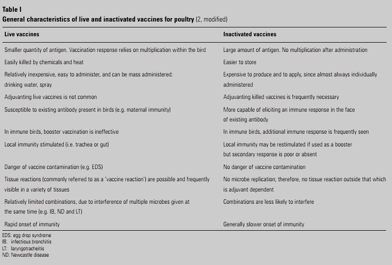
VACCINATION IN POULTRY
Introduction
Poultry are sources of animal protein throughout the world. Moreover, poultry are able to adapt to most geographical areas and conditions, they are not expensive to buy, they have rapid generation time and a high rate of productivity, and they do not require large areas of land.
Vaccines are, in fact, an important component of poultry disease prevention and control worldwide.
Different factors should be considered during vaccination of Poultry:
– the type of poultry production (e.g. commercial or rural)
– the organisation of the industry (e.g. vertical integration)
– the densities of different bird species
– the prevailing disease situation
– vaccine availability
– the use of other vaccines
– the prevalence of other diseases
– the resources available (e.g. manpower and equipment)
– the costs involved.
Herd immunity:
‘Herd immunity’ may be defined as the reduced probability of an individual (bird or flock) becoming infected whenever it is part of a vaccinated population .
Herd immunity is important at two levels:
1)Flock level: if a single bird in a vaccinated flock is not immunised, it has a chance of becoming infected which is inversely proportional to the level of protection achieved by the other vaccinated and immunised birds in the same flock;
2)country/region/compartment level: the higher the prevalence of vaccinated flocks in the vaccination area, the lower the probability of infection in unvaccinated flocks located in the same country/ area/ compartment.
FACTORS WHICH CAN AFFECT THE OUTCOME OF A VACCINATION PROGRAMME
The most important aspects to be considered in improving the organisation of a vaccination programme and achieving the expected outcomes will be briefly illustrated below.
Poultry sector involved
The practical application of poultry vaccines is highly influenced by the characteristics of the poultry producing system in question. Generally speaking, there are two main types of poultry production: industrially reared poultry and rural poultry. The spread of an infectious poultry disease and the measures to be applied for its control, including vaccination, are clearly related to the structure and organisation of the local poultry sector.
Prevailing disease situation
The application of the different vaccination options should be adjusted in diverse conditions according to the local pattern of disease, the level of biosecurity practised in different types of poultry production systems, and the level of challenge for each type of poultry operation. This overall risk assessment should allow for the correct identification of the area/compartment that is to be subjected to vaccination and the optimal vaccination protocol. Furthermore, it is fundamental to monitor the prevalence of infectious agents capable of producing immunosuppression (e.g. infectious bursal disease, infectious anaemia, and Marek’s disease in chickens, and haemorrhagic enteritis in turkeys) and to implement specific vaccination programmes for their control.
Vaccination strategy
Generally speaking, there are three vaccination strategies: routine, emergency and preventive vaccination. Routine vaccination can be the tool of choice in territorial areas where an infectious disease is endemic. Used properly, routine vaccination is effective in reducing mortality and production losses.
Emergency vaccination is an option whenever a new infectious disease is introduced in a previously unaffected country/area/compartment, and the epidemiological situation indicates that there could be massive and rapid spread of infection.
If the disease becomes endemic, the option of applying vaccination on a routine basis can be considered.
Preventive vaccination is a measure that may be applied wherever a high risk of introduction and further spread of a contagious poultry disease has been identified. Preventive vaccination is generally carried out for the prevention of poultry diseases that have a clear impact on the industry. For example, as regards ND(New Castle diseases) control.
Cost/benefit analysis
Before implementing a vaccination programme, an overall cost/benefit analysis should be performed by taking into account the costs of vaccines, vaccine delivery (e.g. labour, equipment), monitoring, laboratory testing, and all other related activities.
Availability of different types of vaccines
Vaccines used in poultry production are classically described as live or inactivated. The availability of different types of vaccines could be one of the major limits to the implementation of effective vaccination programmes. Different types of poultry production (or bird species) or diverse levels of risk require the application of more than one type of vaccine to obtain a high and long-lasting immunological response. As regards ND control, the immune response induced by live ND vaccines increases as their pathogenicity increases.
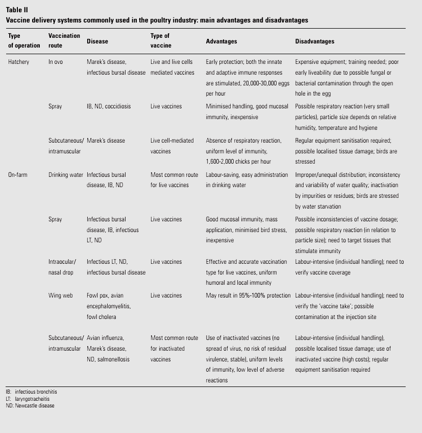
Administration of vaccines
After establishing the type of vaccine to be used, the route, method and frequency of administration must be defined, as well as the proper way to combine all these components in the vaccination programme. Vaccine delivery systems significantly influence the outcome of vaccination. An improper vaccine application is considered one of the most common reasons for vaccination programme failure.
why we should study Agrovet pharmaceuticals in pharmacy?
to gain knowledge about the physiological studies of the animals of our surroundings.
to learn about the diseases and diagnosis of animals.
to study about the similarities and dissimilarities between humans and animal physiology.
to gain knowledge about the diseases that come from animals and humans through pathogens and how to prevent them.
to teach the treatment of disease and invent medicines depending on various factors.
Musculoskeletal problems and treatment of the cattle
Musculoskeletal problems are injuries or pain in the musculoskeletal system, including the joints, ligaments, muscles, nerves, tendons and structures that support limbs, neck and back.
Infectious Pododermatitis (Foot rot)
It is acute and highly infectious disease of cattle characterized by swelling and lameness.
Cause:
Bacteria such as Fusobacterium necrophorum and Bacteroides melaninogenicus.
Symptoms:
1.Pain,Fever,
2.Sudden lameness,
3.Foot will have a foul odor
4.Reduced milk production,
5.Loss of appetite.
Treatment:
1.Early administration of systemic antibiotics,
2.Foot trimming.
Prevention:
1.Environmental hygiene,
2.Promote Drainage.
Spastic Arthritis /Infectious Arthritis
Infectious Arthritis describes the presence of a bacterial , viral or fungal infection in joints.
Cause:
Mainly bacteria, Corynebacterium pyogens and Brucella abortus.
Symptoms:
1.Lameness in one or more joint.
2.Fever.
3.Lymphadenopathy
4.Depression,
5.Anorexia.
Treatment:
1. Antibiotics should be administrated for a minimum 4-6 weeks.
2. NSAIDs can be administrated for pain.
Prevention:
Drainage and flushing is mandatory.
Laminitis
A painful inflammatory condition of the tissues that bond the hoof wall.
Cause:
1.High intake of soluble carbohydrates.
2.Obesity,Concussion.
3.Cushing’s disease
Symptoms:
1.Lameness.
2.Unwilling to stand.
3.Calf crawling on the ground.
Treatment:
No treatment actually. Sometimes Vasodilator and Anti-inflammatory agents can be given.
White Line Disease
A fungal infection of the cattle’s hoof.
Cause:
The exact organism that causes white line disease is not known, but it is known to be caused by bacteria in the soil getting into a weakened spot in the hoof wall which then set up a fungal infection.
Symptoms:
1.Change in the typical light color of the white line to a black or dark gray color.
2.Lameness.
Treatment:
1.By applying Borax or Bleach solution to the affected area of the hoof.
2.Cut away the hoof wall over the affected area.
Degenerative Joint Disease/Osteoarthritis
DJD is a degradation of the articular cartilage.
Cause:
Due to morphological changes in joints.
Symptoms:
1.swelling and deformity of joints.
2.Lameness.
3.Reduced milk production.
4.Have difficulty to stand up and walk.
5.Chronic pain.
Treatment:
1.Use of NSAIDs.
2.Intra articular corticosteroids.
3.IM pentosan polysulfate.
Spastic Syndrome
It is a genetic disease. The condition may progress to posterior paresis or hind limb paralysis.
Cause:
Possibly due to an autosomal dominant gene with incomplete penetrance.
Symptoms:
1.Pain, particularly in feet or joints.
2.Unable to move forward.
3.Stands trembling.
Treatment:
1.Mephenesin (30-40 mg/Kg, for 2-3 days),
2.Phenylbutazone may also have beneficial effects.
Double sole in the cattle
In double sole, one sole is present while a new sole grows beneath.
Cause: Caused by Fusobacterium necrophorum and Bacteroides nodosus
Symptoms: If foreign body penetrate through the corium, pain, unwilling to stand.
Treatment and prevention:
Foreign body should be removed .
Antibiotics should be squeezed into the cavity .
Hairy heel warts
This condition affects most confinement dairies. Lesions typically appear most commonly at the bulb of the heel, but may also be seen at the front of the interdigital cleft . Over 80% of lesions are in the hind foot.
Symptoms:
The term “hairy heel warts” derives from the appearance of hair-like papillary projections of hyperkeratotic skin.
Cause:
Intralesional invasive spirochete organisms resembling Treponema spp. are seen.
Treatment
It is almost impossible to eradicate this condition from a herd. It is only possible to manage it through consistent and persistent application of control methods. Practical options are individual cow treatment (foot wrapping),use of footbaths or spraying lesions with a garden sprayer.
Topical spraying
Add ¼ cup laundry detergent to 2.5 gallon sprayer.
Be careful not to spray udder or milking equipment.


Comments
Post a Comment